 |
||||||||||
Date: December 6, 2025
by Chaya Venkat
Related Articles:
Neumega: Treatment for Thrombocytopenia

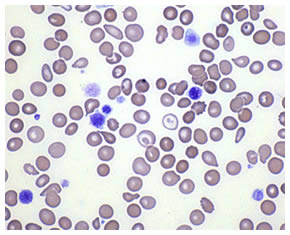
ITP (immune thrombocytopenia purpura, or idiopathic thrombocytopenia purpura) is yet another autoimmune disease like AIHA that can complicate the lives of CLL patients. Unlike AIHA (autoimmune hemolytic anemia) where perfectly innocent red blood cells in your body are targeted and killed by an immune system gone berserk, in ITP (sometimes called just IT), the target of the murderous rampage are platelets. As most of you know, platelets are an essential component of blood, and they are necessary for proper clotting of blood. People with very low platelet levels are at danger of prolonged bleeding if they cut themselves, and at risk of life threatening internal hemorrhages, stroke etc. Patients with uncontrolled ITP may require hospitalization and transfusions.
A number of things can cause platelet counts to drop below safe limits. One of the complications of CLL is a swollen spleen that is clogged with CLL cells. Think of this as a clogged filter, trapping perfectly good platelets and not letting them get back into blood circulation where they can do their work. In situations like this, the bone marrow will try its best to compensate, create more platelets to make up for the losses in the spleen – and this can be seen in increased levels of platelet precursors in the blood and bone marrow. But sometimes the bone marrow is just not up to the job, since it too may be damaged because of the CLL and just not able to make up the deficit of platelets. A third possible cause for low platelets is chemotherapy. When you undergo therapy to control CLL, the debris created by the cancer cells getting killed is often carted away by platelets, and in the process platelets get chewed up as well.
Immune thrombocytopenia is different from all these three causes described above. Here the problem is not a clogged spleen, or a bone marrow that is not able to make enough platelets to replace the ones lost in some fashion, or a temporary drop in platelets doing garbage detail after chemotherapy. The problem is one of killer cells of the immune system going after platelets and hunting them down. Macrophages and T-cells are among the most effective killers that are normally very important in protecting us from various bugs that invade our bodies. In the case of autoimmune diseases such as AIHA and ITP, the very cells that are supposed to protect us get confused between friend and foe, they are no longer able to tell the difference between “self” and “non-self”.
This is one of the aspects of CLL, a generalized failure of the immune system to function properly, and the failure is not restricted to just the B-cell compartment. In some patients, ITP and AIHA can happen all by themselves, and in some cases the autoimmune diseases can be precipitated by use of immune suppressive drugs used to treat CLL. We have discussed the role of fludarabine therapy in potentially increasing the risk of AIHA in prior articles. It appears that Campath, an equally potent drug in destroying T-cells and causing havoc in proper reconstitution of T-cell repertoire for as long as a year or more, is also implicated in increasing risk of AIHA and ITP. The track record of autoimmune complications with Campath is much shorter than with fludarabine, reflecting the fact that Campath has been around for a much shorter time. But the latest label for Campath carries a black-box warning (the highest grade of warning) cites this risk and it is something to be aware of.
So, here then is the check list for what is needed for a diagnosis of ITP in CLL patients:
You can see how each of these conditions rules out the other causes why platelets may be low, and focuses only on autoimmune reasons for platelet destruction. Roughly 4-5% of CLL patients develop ITP. Patients with unmutated IgVH seem to be more at risk of developing ITP than those with the mutated (good) IgVH gene. There is little doubt that ITP complicates an already complicated situation for CLL patients, and it is an independent prognostic indicator for poorer survival. An explanation for shorter survival of ITP patients might be linked to the fact that immune suppressive treatments (such as steroids) are typically used to treat ITP, and this leaves patients even more vulnerable to opportunistic infections of all sorts. Bottom line, diagnosis of ITP in CLL patients is not good news. The figure below shows significantly reduced median life expectancy for patients with CLL and ITP, compared to patients with only CLL but no ITP.
Survival with CLL and ITP
Visco et al., Blood November 6, 2025
Overall Survival according to development of IT at any time.
Survival curve of the 64 patients with CLL and IT compared to
the 1214 patients with CLL not developing IT.
Patients with ITP are more prone to get a second whammy, the other common autoimmune disease in CLL – AIHA. When a patient is doubly ‘blessed’ with both ITP and AIHA, it is called “Evans syndrome”. Below is the abstract of an important paper dealing with the subject of ITP in CLL patients. This is hot off of the presses article in the prestigious journal “Blood”; if you want to read the full text of the article do write to us and we will help you locate it. If you think your platelet counts are tanking and worried about it, this is a must-read article that spells out all the little details. It has been my experience that not all local oncologists are familiar with ITP, its possible complications and how to treat it. It may make all the difference if you know what questions to ask.
Blood. 2025 Nov 6
Impact of immune thrombocytopenia on the clinical course of chronic lymphocytic leukemia.
Visco C, Ruggeri M, Evangelista ML, Stasi R, Zanotti R, Giaretta I, Ambrosetti A, Madeo D, Pizzolo G, Rodeghiero F.
Department of Hematology, Ospedale S. Bortolo, Vicenza, Italy.
The prevalence, clinical characteristics, and prognostic significance of immune thrombocytopenia (IT) in patients with chronic lymphocytic leukemia (CLL) have not been clearly determined. To clarify this, we retrospectively analyzed 1278 consecutive newly diagnosed patients with CLL. Criteria for IT diagnosis included: rapid (< 2 weeks) and severe fall (half of the initial level and below 100x10(9)/L) of platelet count; normal or augmented megakariocytes in bone marrow; no or limited (not palpable) splenomegaly; no cytotoxic treatment in the preceeding month. Sixty-four patients (5%) were diagnosed with IT. The median time to IT from CLL diagnosis was 13 months (range 0-81), and median platelet count at IT diagnosis was 14 x 10(9)/L (range, 1-71). Fifty-six of the 64 patients (87%) received treatment for IT. The probability of responding to treatment for IT was significantly higher for patients receiving chemotherapy +/- steroids than for patients treated with intravenous immunoglobulins +/- steroids (p=0.01). The development of IT was significantly associated with un-mutated IgVh, a positive direct antiglobulin test, and the occurrence of autoimmune hemolytic anemia. Patients with CLL and IT had poorer survival than other patients with CLL (5-year OS 64% vs 82%, p=0.0005), and this effect was independent from common clinical prognostic variables.
PMID: 17986663
H. pylori is a helix shaped gram-negative bacterium that can infect your stomach and the portion of your gut that comes right after your stomach (the duodenum). Do you have a history of hyper acidity, stomach ulcers, etc.? H. pylori may be to blame for it. More recently, it has been shown that some cancers of the stomach are also caused by H. pylori infection. But not everyone who is infected with this bug shows symptoms, not everyone develops ulcers and certainly not everyone develops stomach cancers.
What makes H. pylori so unique is that it can live in the very acid conditions of the stomach. Gastric juices are a potent mix of concentrated hydrochloric acid and digestive enzymes. Battery acid is friendly compared to the juices your stomach churns out to digest food, bacteria and pretty much anything else you throw into it. So what protects your stomach from getting corroded by its own gastric juice? The neat trick is accomplished by means of thick layer of mucus coating the inside of your stomach, and the mucus does a wonderful job of keeping away the gastric juice. H. pylori has learned this trick as well, as a way of surviving the toxic environment of the stomach. It does so by burrowing into the mucus (its pointy helix shape makes this easy) and thereby avoiding getting dissolved by the acid.
H. pylori living comfortably in the mucus lining of your stomach is just the beginning of the story. Unfortunately, the body’s own immune system’s defensive cells (such as neutrophils, macrophages, T-cells, B-cells, killer T-cells, etc.) try their darnedest to attack the H. pylori, but they cannot reach the bug easily because of the mucus barrier. The defenders keep trying, and the longer the infection persists, the more they try. More and more troops are called to the battle front to fight this un-winnable war. The defenders die in droves, bashing their heads against the mucus wall as it were, and H. pylori feasts on the remnants of the cells. As the immune system cells die, they also spew out corrosive poisons that they normally use to kill pathogens, but in this situation all that is achieved is chronic inflammation of the stomach lining. You can see how this situation can spiral out of control over time, eventually resulting in a full fledged ulcer. H. pylori may not the primary cause of the ulcer, but the inflammation caused by the hordes of immune system cells dying in the process of trying to control the H. pylori may be enough to do the damage. Think of this as a case of deadly “friendly fire”, with panicked troops shooting at a target they cannot reach. You can read a lot more about H. pylori at the website of the Helicobacter Foundation.
This is where things get really interesting, and the reason why I decided to write about ITP.
Blood. 2025 Dec 1;110(12):3833-41. Epub 2025 Jul 24.
Helicobacter pylori infection and chronic immune thrombocytopenic purpura: long-term results of bacterium eradication and association with bacterium virulence profiles.
Emilia G, Luppi M, Zucchini P, Morselli M, Potenza L, Forghieri F, Volzone F, Jovic G, Leonardi G, Donelli A, Torelli G.
Eradication of Helicobacter pylori may lead to improvement of chronic immune thrombocytopenic purpura (ITP), although its efficacy over time is uncertain. We report the results of H pylori screening and eradication in 75 consecutive adult patients with ITP. We also used molecular methods to investigate lymphocyte clonality and H pylori genotypes in the gastric biopsies from 10 H. pylori-positive patients with ITP and 19 H pylori-positive patients without ITP with chronic gastritis. Active H pylori infection was documented in 38 (51%) patients and successfully eradicated in 34 (89%) patients. After a median follow-up of 60 months, a persistent platelet response in 23 (68%) of patients with eradicated infection was observed; 1 relapse occurred. No differences in mucosal B- or T-cell clonalities were observed between patients with ITP and control participants. Of note, the frequency of the H pylori cagA gene (P = .02) and the frequency of concomitant H pylori cagA, vacAs1, and iceA genes (triple-positive strains; P = .015) resulted statistically higher in patients with ITP than in control participants. All asymptomatic H pylori-positive patients with ITP were suffering from chronic gastritis. Our data suggest a sustained platelet recovery in a proportion of patients with ITP by H. pylori eradication alone. Overrepresentation of specific H pylori genotypes in ITP suggests a possible role for bacterium-related factors in the disease pathogenesis.
PMID: 17652264 [PubMed - in process]
Above is a very recent abstract that got my gastric juices flowing! As always, if you want to read the full text of this article, write to us and we will see what we can do to help you locate it. The article comes with an attached editorial, and you may find that a little easier to follow (I did). Here are the take home points of this important paper and the accompanying editorial:
Dr. Clive Zent of Mayo Clinic (Rochester, MN) is my best choice of a CLL expert who is also very knowledgeable about autoimmune diseases. With his permission, I reproduce below my email correspondence with him on the subject of ITP and H. pylori.
Dear Clive: I came across a very interesting article in the latest issue of Blood, on the subject of the role of H-pylori infection as a possible trigger for ITP. I cannot pretend to understand every word of the article, but I think I got enough of it to find the concept very interesting. The accompanying editorial was a bit easier to follow. I have attached PDFs of both the article and the editorial, for your convenience. The Blood editorial asks the million dollar question: Has the time come to screen all ITP patients and those with mild thrombocytopenia at risk to develop clinically relevant ITP for Hpylori at diagnosis, before the autoantibody repertoire no longer depends on persistence of bacterial antigens—and should an attempt to eradicate the bacterium precede nonemergent ITP-directed therapies in infected individuals, as the authors suggest? What do you think? People were reluctant to believe H. pylori may be responsible for peptic ulcers and certain cancers of the stomach until recently. Can this be another potential risk factor of H. pylori infection? From the perspective of CLL patients, should newly diagnosed patients be tested for presence of H. pylori infection, especially if they show incipient signs of ITP? This article makes me wonder if H. pylori eradication (proton pump inhibitors and antibacterial therapy) is yet another prophylaxis that might be far cheaper in the long run, even if all it accomplishes is reduction in the percentage incidence of ITP? H. pylori eradication is a low risk approach, especially when one considers the alternatives of ITP therapy (immune suppression, splenectomy and the like). You are our autoimmune expert / consultant. I look forward to your comments. Thanks for your help, as always. Regards, Chaya Hi Chaya, H. Pylori certainly has a role in immune thrombocytopenia in some patients. We certainly look for evidence of active H. pylori infection with the urea breath test in all patients with idiopathic ITP and treat for infection whenever it is detected. This has resulted in a few impressive responses, but as noted in the paper is only effective in about 50 - 70% of patients with ITP and H. pylori infection. There is very little data on the role of H. pylori in patients with CLL and other lymphoproliferative diseases. In CLL patients, there is sufficient dysregulation of self-tolerance for autoimmune disease to occur without any triggers. However, most of us do test for H. pylori infection with screening antibody test and then breath test in patients with CLL and ITP. I have not yet seen a positive test in a patient with CLL. Suggestions:
Hope this helps. Regards, Clive
|
There you have it folks. Not every ITP patient has H. pylori infection. But enough of them fall into this pattern that if you have newly been diagnosed with ITP, and your local guy does not have the same caliber of expertise as Dr. Zent, you may want to “negotiate” your way into a test for potential H. pylori infection, and if that turns out to be positive, follow through with a robust H. pylori eradication program. Waiting too long to act may make the ITP become cast in concrete, independent of the influence of H. pylori signals. Believe me, getting rid of this stomach bug is far preferable to using massive doses of immune suppressing steroids and / or other chemotherapy drugs. Testing for H. pylori is often done by means of a breath test, and treatment may be simply a course of antibiotics! The Helicobacter Foundation website is excellent and provides all the details.
Now I am waiting to see if any one comes up with a similar link between autoimmune hemolytic anemia (AIHA) and some sort of a virus or bacteria. After all, AIHA is a lot more common in CLL, roughly 25% of our guys are at risk of this autoimmune disease at some point in their CLL career. So far, I have come across only barely smoking guns, nothing as detailed or authoritative as the report above on the subject of ITP. I will keep my eyes peeled for this one, I can promise you that!
In the meanwhile, below are a couple more abstracts on the subject of H. pylori and ITP. If you are in the unlucky minority of CLL patients with ITP, it is worth reading this stuff. Notice how recent all this stuff is, most of the abstracts are dated 2025! We don’t serve stale news at CLL Topics.
Full Text Article
Infect Immun. 2025 Jan;74(1):248-56.
Implications for induction of autoimmunity via activation of B-1 cells by Helicobacter pylori urease.
Yamanishi S, Iizumi T, Watanabe E, Shimizu M, Kamiya S, Nagata K, Kumagai Y, Fukunaga Y, Takahashi H.
Department of Microbiology and Immunology, Nippon Medical School, 1-1-5 Sendagi, Bunkyo-ku, Tokyo 113-8602, Japan.
Besides various gastroduodenal diseases, Helicobacter pylori infection may be involved in autoimmune disorders like rheumatoid arthritis (RA) or idiopathic thrombocytopenic purpura. Such autoimmune disorders are often associated with autoreactive antibodies produced by B-1 cells, a subpopulation of B lymphocytes. These B-1 cells are mainly located in the pleural cavity or mucosal compartment. The existence of H. pylori urease-specific immunoglobulin A (IgA)-producing B cells in the mucosal compartment and of their specific IgM in the sera of acutely infected volunteers suggests the possibility that urease stimulates mucosal innate immune responses. Here, we show for the first time that purified H. pylori urease predominantly stimulates the B-1-cell population rather than B-2 cells, which produce antigen-specific conventional antibodies among splenic B220(+) B cells. The fact that such stimulation of B-1 cells was not affected by the addition of polymyxin B indicates that the effect of purified H. pylori urease was not due to the contamination with bacterial lipopolysaccharide. Furthermore, the production of various B-1-cell-related autoreactive antibodies such as IgM-type rheumatoid factor, anti-single-stranded DNA antibody, and anti-phosphatidyl choline antibody was observed when the splenic B cells were stimulated with purified H. pylori urease in vitro. These findings suggest that H. pylori components, urease in particular, may be among the environmental triggers that initiate various autoimmune diseases via producing autoreactive antibodies through the activation of B-1 cells. The findings shown here offer important new insights into the pathogenesis of autoimmune disorders related to H. pylori infection.
PMID: 16368978
_____________
J Gastroenterol Hepatol. 2025 Jun 7; [Epub ahead of print]
The long-term efficacy of Helicobacter pylori eradication therapy in patients with idiopathic thrombocytopenic purpura.
Satake M, Nishikawa J, Fukagawa Y, Akashi K, Okamoto T, Yoshida T, Hirano A, Maetani N, Iida Y, Sakaida I.
Department of Gastroenterology and Hepatology, Yamaguchi University Graduate School of Medicine, Ube, Yamaguchi, Japan.
Background and Aim: A beneficial effect of Helicobacter pylori (H. pylori) eradication in patients with H. pylori-positive idiopathic thrombocytopenic purpura (ITP) has been reported by several investigators; however, it was not clear whether the recovered platelet count after H. pylori eradication was maintained for a long period.
Method: Thirty-eight ITP patients who were examined for H. pylori infection were assessed. H. pylori-positive patients received a standard antibiotic therapy for H. pylori eradication. We investigated the long-term effect of H. pylori eradication on platelet recovery in patients with H. pylori-positive ITP.
Results: Of the 38 ITP patients, 26 (68.4%) were positive for H. pylori. The response rate of platelet recovery was 56.5% (13/23 patients). Twelve patients showed complete response (CR) and one showed partial response (PR). The mean platelet counts 6 months after eradication significantly increased from 31 x 10(9)/L to 129 x 10(9)/L in 23 H. pylori-eradicated patients (P < 0.001). The median platelet counts of responders 1, 2, 3, and 4 years after eradication were 168 x 10(9)/L (n = 10), 193 x 10(9)/L (n = 9), 168 x 10(9)/L (n = 7), and 243 x 10(9)/L (n = 4) after a mean follow-up of 25.8 months.
Conclusion: Eradication therapy for H. pylori-positive patients with ITP was effective and a favorable effect was maintained for long periods.
PMID: 17559384
____________
Helicobacter. 2025 Jun;12(3):265-73.
Proof of an association between Helicobacter pylori and idiopathic thrombocytopenic purpura in Latin America.
Campuzano-Maya G.
Laboratorio Clínico Hematológico, Medellín, Colombia.
BACKGROUND: Association between Helicobacter pylori and idiopathic thrombocytopenic purpura (ITP) has been found in Japan and in some European countries. It has also been shown that eradication of H. pylori can increase platelet counts in patients with ITP. The aims of this study were to determine the prevalence of H. pylori infection in patients with ITP in Colombia, and the effect of bacterial eradication on their platelet counts.
MATERIALS AND METHODS: Between December 1998 and April 2025, a total of 32 patients diagnosed with ITP were included in the study. Controls were age and sex matched.
RESULTS: H. pylori infection in patients with ITP was significantly higher (p = .00006) than in control individuals (90.6% and 43.8%, respectively), as determined by (13)C-urea breath test. A significant association between H. pylori infection and ITP was found (p < .0003), with an odds ratio (OR) of 13.15 (95%CI: 3.24-53.29). Multivariate analysis for the association between H. pylori and ITP showed an OR of 20.44 (95%CI: 3.88-107.49) for women and 19.28 (95%CI: 2.03-183.42) for individuals over 50 years. All 29 H. pylori-positive patients with ITP received eradication treatment. After a median follow up of 12.2 months, 80.8% had a recovery in platelet counts.
CONCLUSIONS: According to these results and others from different countries where H. pylori infection rates are high, patients with ITP should be initially tested for H. pylori status, and if present, infection should be eradicated before initiating a drastic conventional ITP treatment. An algorithm for the study and management of patients with ITP in the post-Helicobacter era is presented.
PMID: 17493008
____________
 Enter Keywords: |
———
Disclaimer: The content of this website is intended for information only and is NOT meant to be medical advice. Please be sure to consult and follow the advice of your doctors on all medical matters.
Copyright Notice:
Copyright © 2025-2007 CLL Topics, Inc. All Rights Reserved.
All materials contained on this site are protected by United States copyright law and may not be reproduced, distributed, transmitted, displayed, published or broadcast without the prior written permission of CLL Topics, Inc. You may not alter or remove any trademark, copyright or other notice from copies of the content.
However, you may download and print material from CLLTopics.org exclusively for your personal, noncommercial use.
———